

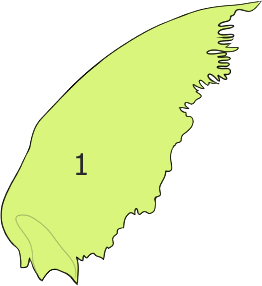


1. Frontal Bone: Composed of the squamous and orbital parts. The squamous portion encompasses the forehead. It is vertical and relatively flat. The orbital portion includes the upper sections of the orbital and nasal cavaties. Notable features include the supraorbital ridges (browridge) and the anterior portions of the temporal lines.
2. Sphenoid Bone: Seated in front of the basilar part of the occipital bone. The median portion contains the body of the sphenoid bone, which encompasses the sella turcica and sphenoidal sinuses. The wings are comprised of two larger wings attached to the lateral sides of the body and two smaller wings on the anterior sides of the body. Articulates with the frontal, ethmoid, temporal, parietal, vomer, and occipital bones to form the middle-posterior section of the orbits.
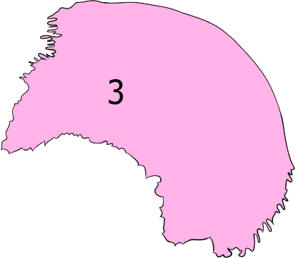
3. Parietal Bone: Relatively large compared to other bones of the skull. Form the sides and superior areas of the cranium. Identifiable by the presence of meningeal grooves on the inside surface, which branch out posteriorly towards the back of the skull.
4. Ethmoid Bone: Divides nasal cavity and brain. Found posteriorly between the orbits, aligned with the nasal bones. Articulates with the frontal, sphenoid, nasal, maxillae, palatine, vomer, and lacrimal bones.
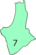
7. Zygomatic Bone: Paired bones which form the cheeks (cheekbones), as well as the lateral wall and floors of the orbits. Articulates with the frontal, sphenoid, temporal, and maxillary bones.
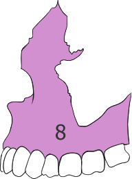
8. Maxilla: Upper jawbone. Formed by two paired maxillary bones which fuse in the center. Contains the maxillary sinus and anterior portion of the hard palate.
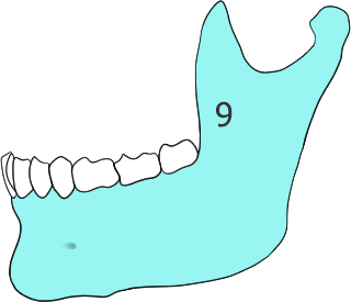
9. Mandible: Bottom jawbone. Articulates with the temporal bones.
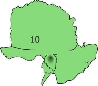
10. Temporal Bone: Form the temples and contain components of the ears. Identified easily by temporal line (which connects with the portion of the ridge seen on the frontal bone) as well as a long, narrow styloid process, though this often breaks off.
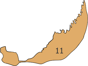
11. Occipital Bone: Bone which forms the posterior base of skull. Identifiable by large hole known as the foramen magnum (facillitates connection of medulla oblongata and spinal cord), as well as a raised cross-shape on the interior portion known as the cruciform eminence.
2. Sphenoid Bone: Seated in front of the basilar part of the occipital bone. The median portion contains the body of the sphenoid bone, which encompasses the sella turcica and sphenoidal sinuses. The wings are comprised of two larger wings attached to the lateral sides of the body and two smaller wings on the anterior sides of the body. Articulates with the frontal, ethmoid, temporal, parietal, vomer, and occipital bones to form the middle-posterior section of the orbits.
3. Parietal Bone: Relatively large compared to other bones of the skull. Form the sides and superior areas of the cranium. Identifiable by the presence of meningeal grooves on the inside surface, which branch out posteriorly towards the back of the skull.
4. Ethmoid Bone: Divides nasal cavity and brain. Found posteriorly between the orbits, aligned with the nasal bones. Articulates with the frontal, sphenoid, nasal, maxillae, palatine, vomer, and lacrimal bones.
5. Lacrimal Bone: Two small paired bones found on the medial wall of the orbit, near the front. Articulates with the frontal, ethmoid, maxillae, and inferior nasal conchae.
6. Nasal Bone: Two paired bones which form the bridge of the nose. Articulate with the frontal, ethmoid, and maxillae bones. 7. Zygomatic Bone: Paired bones which form the cheeks (cheekbones), as well as the lateral wall and floors of the orbits. Articulates with the frontal, sphenoid, temporal, and maxillary bones.
8. Maxilla: Upper jawbone. Formed by two paired maxillary bones which fuse in the center. Contains the maxillary sinus and anterior portion of the hard palate.
9. Mandible: Bottom jawbone. Articulates with the temporal bones.
10. Temporal Bone: Form the temples and contain components of the ears. Identified easily by temporal line (which connects with the portion of the ridge seen on the frontal bone) as well as a long, narrow styloid process, though this often breaks off.
11. Occipital Bone: Bone which forms the posterior base of skull. Identifiable by large hole known as the foramen magnum (facillitates connection of medulla oblongata and spinal cord), as well as a raised cross-shape on the interior portion known as the cruciform eminence.